DENTAL XRAYS! WHATS THE RIGHT ONE FOR ME
What are dental xrays used for
Dental xrays were created for one reason, to examine further what the naked eye can not see. This means that we can see whats going on inside your teeth your body. Xrays traditionally are best used to check the hard tissue meaning bones and teeth. The imaging provided serves as a very helpful diagnostic tool for dentists.
Dixhital vs Analog xrays
Digital graphics have long since supplanted analog graphics, which were commonly employed in Albania and used Kodak film. The explanation is quite straightforward: digital technology requires 20 times less radiation to expose digital film. What benefits do digital graphics offer?
Benefits of digital dental xrays
Compared to graphite film, the radiation dosage utilized is twenty times lower.
In contrast to Kodak film, the image is crisper and the dentist can electronically adjust it to view more precise image details.
Instead of using single-use Kodak film, only an electronic sensor is utilized, which lowers recycling costs and pollution to the environment.
They can be emailed to you for free as a record and remain on the computer for future treatments, making them easier to save.
Most used dental xrays
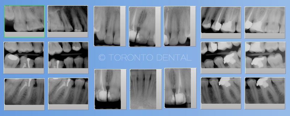
The most xommon xrays in dentistry are PA’s or Periapical xrays, which are used to diagnose small fillings, remove nerves from teeth, diagnose filling difficulties in general, and more.
The American & Canadian Dental Associations advises:
FMX full mouth xrays are routinely performed every two years and consist of eighteen compact digital xrays that offer the most accurate details for the majority of dental disorders.
Panoramic xray
These types are not recomended as often anymore due lesser accuracy compared to the new technology like 3d scaner. Is is mostly used to have a general 2d image of the dentition but it misses the acuracy and the resolution to see better the possible problems.
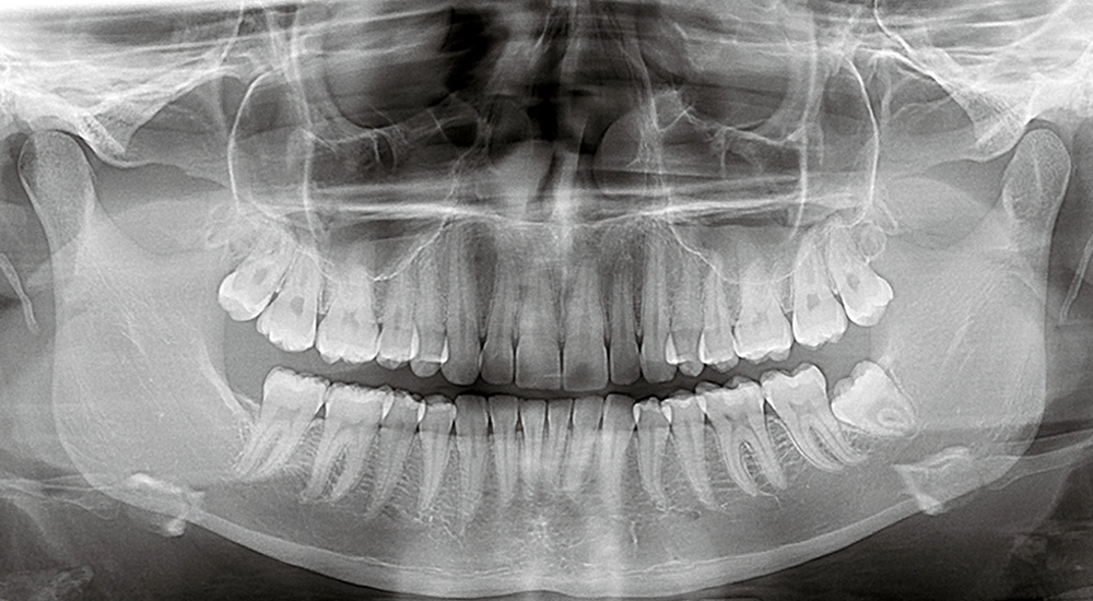
- To see the position of the wisdom teeth which cannot be captured with small xrays
To see the development of children’s teeth from the age of 1 to 13 years. For further factors connected to the kind of care your dentist determines
For those who have trouble expanding their mouth wide or experience acid reflux when they put objects in their mouths
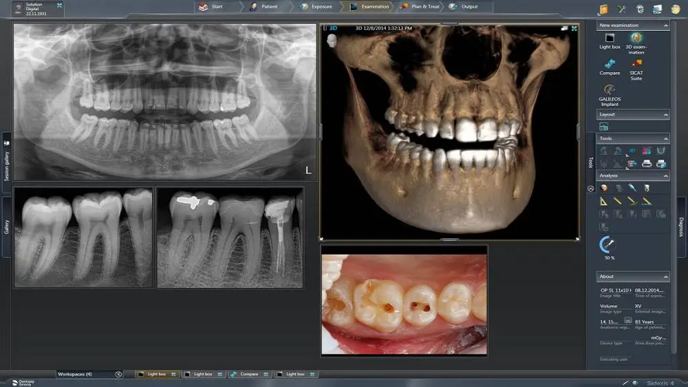
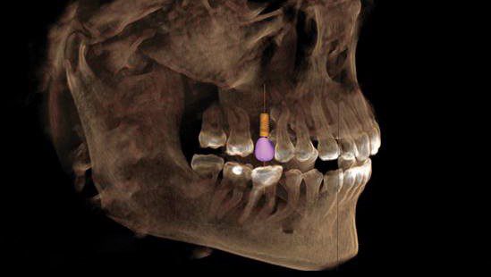
3D dental implant scaners
Enhanced Accuracy: By producing extremely accurate digital representations of the patient’s oral anatomy, 3D Tac scanners provide exact diagnosis and treatment planning. More accurately than with conventional techniques, the detailed images generated by the scanner assist dentists in identifying problems including cavities, bone density, and anatomical features.
Enhanced Patient Comfort: 3D Tac scanners employ non-invasive technology to swiftly and painlessly capture digital impressions, in contrast to traditional dental impressions that require patients to bite down on unpleasant trays loaded with impression material. Patients no longer experience the same level of discomfort or anxiety from classic impression-taking techniques.
Streamlined Workflow: By doing away with the need for actual impressions, which frequently take longer to prepare, ship, and process, 3D Tac scanners simplify the dental workflow. Digital scans allow dentists to view and share patient data instantly with experts or dental laboratories, speeding up treatment and minimizing patient chair time.
Comprehensive Visualization: By creating precise, three-dimensional images of a patient’s oral cavity, 3D Tac scanners give dentists a thorough understanding of the teeth, gums, surrounding tissues, and bone structure. Better treatment planning and communication amongst dental experts are made possible by this, which improves treatment outcomes and efficiency.